What is CBA?
A guide for families
This guide explains what to expect if your baby or child has been diagnosed with a congenital bronchial atresia (CBA).
Medically reviewed by Marisa Schwab, MD
Written by Emily Lake, PhD
Last updated 05/04/2026
What is congenital bronchial atresia?
Congenital bronchial atresia (CBA) is one of the rarer types of congenital lung malformation. It occurs when part of an airway (bronchus) in the developing baby's lung does not form properly, creating a blockage that prevents air from moving in and out of that section of lung normally. CBA is often discovered during a routine prenatal ultrasound, though it is sometimes found after birth. CBA can occur on its own or alongside other lung malformations.
CBA develops happens very early during pregnancy, sometime before the 10th week of gestation. The result is that a small area of lung cannot communicate properly with the rest of the airways. Because air can’t move in and out normally, that area of lung may become over-inflated. On imaging, it may appear as a bright, cystic or air-filled area.
CBA can affect one small segment or an entire lung lobe. It most commonly affects the upper lobes, but it can occur in any part of the lung.
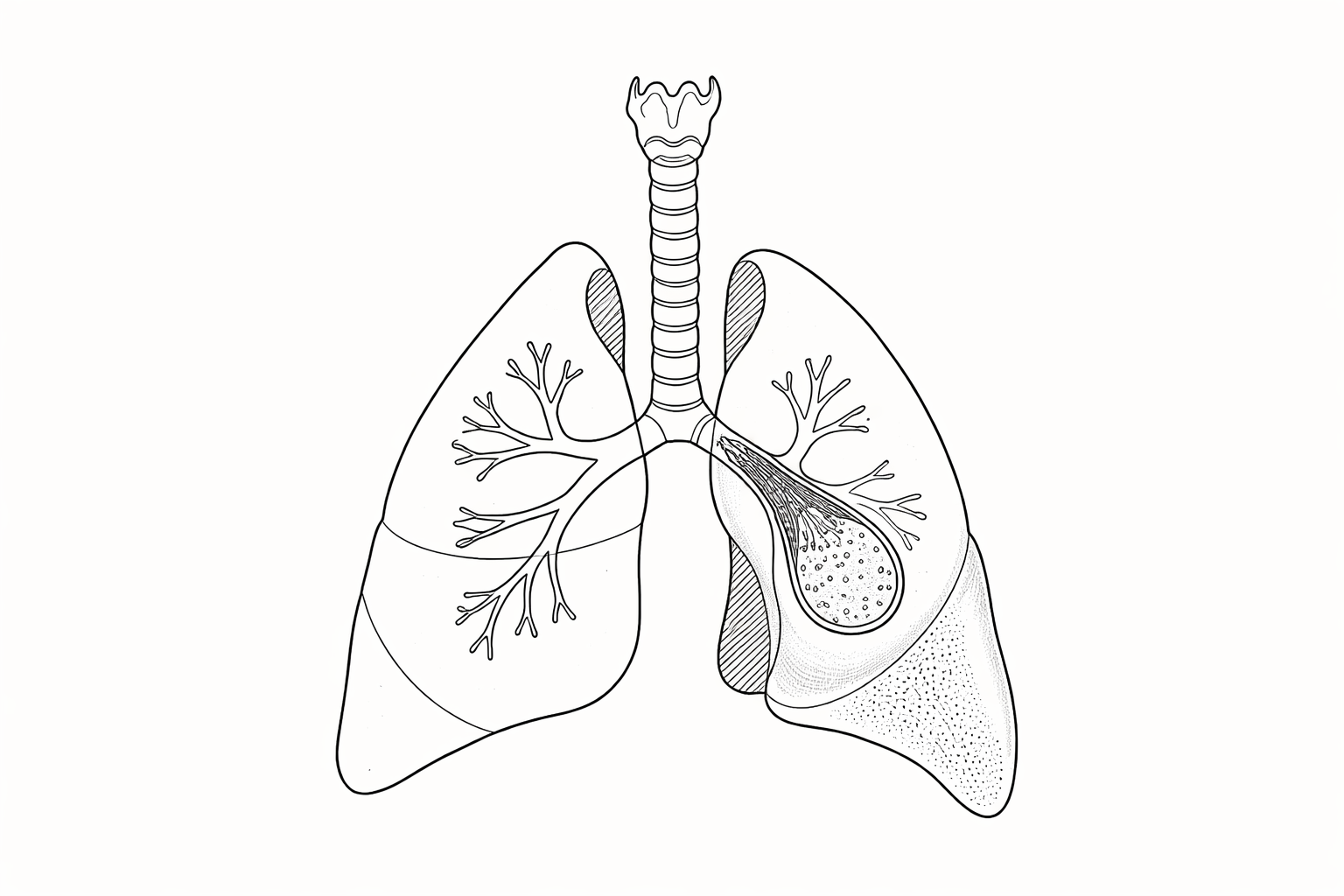
A sketch of the lungs showing congenital bronchial atresia.
CPAM vs CBA
Both are congenital lung malformations involving an area of lung that didn't develop normally in early pregnancy. In CBA, the bronchus (airway) failed to form properly, creating an obstruction. In CPAM, abnormal tissue forms a cystic or solid mass within the lung.
What causes CBA?
The cause of CBA is unknown. Nothing a mother did or didn’t do in pregnancy caused it. These are random malformations that develop in the lungs very early during pregnancy, sometime before the 10th week of gestation.
Diagnosis
CBA is normally found during a routine prenatal ultrasound scan, usually around 20 weeks of pregnancy. The scan technician commonly notices a bright white patch in the baby’s lungs which suggests an abnormal area of tissue. There may be signs that the lesion is pushing on other organs, such as shifting the heart to the other side of the baby’s chest (mediastinal shift). There is also possible flattening of the diaphragm on the side affected by the CBA lesion. Higher risk cases are very uncommon with bronchial atresia.
It can be difficult to distinguish CBA from CPAM before birth because these lesions can look very similar on prenatal imaging. It’s commonly only until a CT scan is done after birth that the medical team will have a good idea of whether this is really CBA or a different type of congenital lung lesion. The CT scan will also give a better idea of the location and characteristics of the CBA. An accurate diagnosis is usually made only after surgical evaluation.
How rare is CBA?
Congenital lung malformations, including CBAs, are uncommon. Estimates for all lesions are 1 in 2,500 live births. CBA is rarer than CPAM and BPS, although it is commonly found as a “hybrid lesion”, meaning for example that CBA and CPAM are both present in the lung tissue. Currently, approximately 70% of babies with a CLM are diagnosed before birth.
Cancer risk and CBA
There is no known pathway for CBA to develop into cancer. There may be an increased risk of cancer with CPAM, and some patients have hybrid lesions, meaning both CBA and CPAM. More research is needed to truly understand the relationship between CPAM and cancer.
Prognosis
Bronchial atresia is one of the rarest congenital lung malformations, but it is also one of the least likely to cause serious problems. While it may look dramatic on prenatal imaging, most babies with CBA do extremely well. For the majority of babies without hydrops during pregnancy, survival is near 100% and long-term outcomes are excellent.
Management and treatment
There are two main paths for bronchial atresia management and treatment. Which one you choose depends on the specifics of your baby’s lung lesion, the advice of your medical team, where in the world you are located, and what you feel is best for your child.
Elective surgical removal (most often a thoracoscopic or open lobectomy) in later infancy— normally around 3–6 months—even if a child has no symptoms. This is the common treatment path in the USA and means that the surgeon will remove the entire affected lung lobe. The reason this is recommended by most doctors in the USA is to prevent repeated and severe infections (such as pneumonia) for the rest of the patient’s life and make the diagnosis certain.
The recovery time depends on whether the operation is done via the open or minimally invasive technique. After a thoracoscopic lobectomy, babies usually recover quickly and spend one or two days in the hospital. The remaining lung will expand and grow to compensate for the removed lobe, and most children will have the same lung function as someone who didn’t have lung surgery as a baby. They’ll be able to do all the activities and sports they want when they grow up.Careful observation (“watchful waiting”) with scheduled check-ins and regular imaging. This is the common treatment path for patients with asymptomatic CBA in Canada, Australia, and many European countries. If symptoms develop—recurrent chest infections, wheezing not explained by common causes, or a spontaneous pneumothorax—surgery is then usually recommended.